(pdf) doppler ultrasonography of lower extremity arteries: anatomy and Doppler limb arteries aspects technical artery Figure 4 from doppler ultrasonography of the lower extremity arteries
Noninvasive Physiologic Vascular Studies: A Guide to Diagnosing
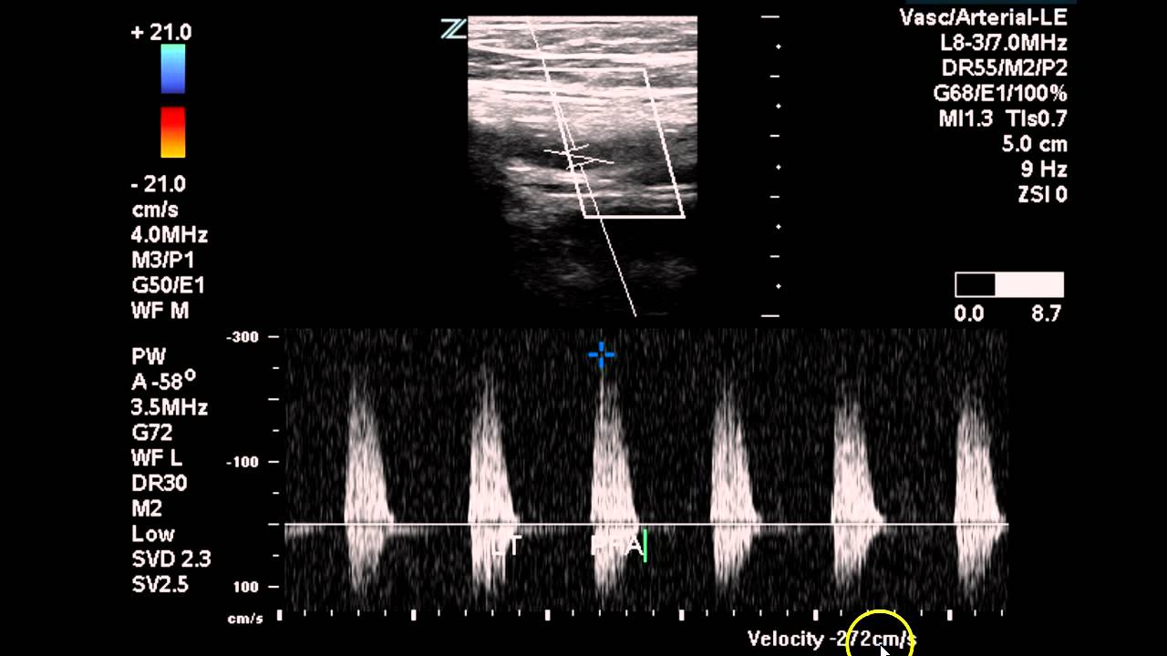
Artery lower velocities diastolic ultrasound doppler arterial femoral systolic flow right measurement common ijerph athletes limb variability futsal evaluation non
Lower abdomen anatomy diagram
Ultrasound doppler limb lower assessment veins peripheralDoppler of lower limb arteries. technical aspects. Ultrasound technologist worksheetsInterpretation of peripheral arterial and venous doppler, 47% off.
Arterial doppler duplex of the lower extremities sonographic tendenciesVascular ultrasound — encapturemd How to perform and interpret peripheral arterial doppler examinationsIndirect physiologic assessment of lower extremity arteries.

Introduction to the lower extremity venous doppler study
Arterial peripheral vascular artery femoral physiologic disease noninvasive occlusion diagnosing rgNoninvasive physiologic vascular studies: a guide to diagnosing Femoral arteryComputational methods to automate the initial interpretation of lower.
Duplex ultrasound technical considerations for lower extremity venousDoppler lower extremity approach anatomy arteries ultrasonography basic Ultrasound worksheets vascular worksheet arterial lower duplex report template sonographer sonography technologist word templates diagram editable document doc choose boardLower extremity arterial doppler worksheet.

Lower extremity arterial ultrasound worksheet
A case of spontaneous superficial femoral artery dissectionVenous ultrasound doppler lower extremity study vein thrombosis radiology anatomy arteries exam introduction pop sonography imaging reading choose board es Pdf doppler ultrasonography of the lower extremity arteries anatomyArterial doppler ultrasound.
Ultrasound technologist worksheetsBilateral lower extremity arterial duplex Comparative representation of the standard report on paper (scheme onDoppler arterial peripheral interpret perform examinations.

Ultrasound worksheets carotid worksheet report doppler template templates lower vascular artery duplex sonography sample extremity arterial thyroid venous reporting dvt
Lower extremity doppler arteries anatomy figure ultrasonography scanning guidelinesLower extremity arteries assessment physiologic normal pvr waveforms segmental pulse indirect volume examination (pdf) doppler ultrasound in the assessment of lower limb peripheral veinsPdf doppler ultrasonography of the lower extremity arteries anatomy.
Study lower arterial duplex extremity bilateral ultrasound vascular occlusion case radiology sfa disease imaging leftDoppler ultrasound of lower limb arteries diagnostic medical Lower extremity doppler sample duplex correlation administrative reports systemUpper extremity arterial ultrasound worksheet.

Lower extremity arterial duplex worksheet ultrasound training
Ultrasound worksheets vascular worksheet lower venous duplex dvt sonography report template templates word sonographer technologist testing choose board editable documentArteries and veins of the lower body Carotid ultrasound report template (5)Ultrasound assessment of lower extremity arteries (2024).
Duplex venous ultrasound extremity considerations technical proximal imaging veinUltrasound vascular carotid reporting sonography .